
By Khalil Al-Salem M.D FRCS, FICO
Quick tips on the eyelid position
- The upper lid margin lies 1–2 mm below the upper corneal limbus.
- The lower lid margin is at the level of the lower corneal limbus.
- The vertical palpebral fissure measures 7–12 mm.
- The upper lid peak lies just nasal to the center of the pupil.
- The adult upper lid crease lies 7–10 mm above the lid margin on down gaze.
- The lateral angle between the two lids is about 2 mm higher than the rounded medial angle.
Blood supply of the eyelid

- Dual anastomoses of the lateral and medial palpebral arteries in each lid.
- Anastomoses with the facial arterial networks.
- the venous system of the eyes has a direct communication with the venous system of the brain (cavernous sinus)
Lymphatic drainage

- The lateral two-thirds of the lids drain to the superficial parotid nodes.
- The medial lids drain to the submandibular nodes.
- tip: the points in violet are important point during the examination of patients with Adenoviral infection. usually the lymphnodes are big and tender.
innervation of the eyelid

- innervation of the eyelid comes from the 5th cranial nerve.
- the following picture shows the dermatomes.
muscles of the eyelid

- The elliptical orbicularis oculi muscle consists of an orbital
part which extends onto the cheek and temporal area, a
palpebral part which is divided into a preseptal, pretarsal and
ciliary parts, and a lacrimal part which extends into the
posterior fascia of the lacrimal sac. - Contraction has a purse-string effect, closing the lids and
drawing the muscle medially. - The lacrimal part dilates the lacrimal sac, contributing to the
lacrimal pump mechanism.
Upper lid retractors
- The striated levator palpebrae superioris (LPS) muscle is innervated by the oculomotor nerve, and has a common origin with the superior rectus muscle. Anteriorly, it becomes the levator aponeurosis as it passes anterior to Whitnall ligament, and inserts into the anterior tarsal surface. Medial and lateral extensions, or horns, insert into the periostium of Whitnall’s tubercle laterally, and into the medial canthal tendon medially. A few anterior fibres pass through the orbicularis to form the skin crease. Contraction of LPS produces 12–20 mm of upper lid elevation.
- The sympathetically innervated Müller’s muscle arises from the posterior surface of the levator muscle, and inserts into the superior border of the tarsus. Contraction elevates the lid by up to 2 mm.
Lower lid retractors
- Arise as the capsulopalpebral head of the inferior rectus, passing forward as a fascial sheet which envelops the inferior oblique muscle and inserts into the inferior tarsus.
- A sympathetically innervated portion lies immediately posterior to the aponeurotic portion, arising from the fascial sheath of the inferior rectus muscle and inserting into the inferior tarsus.
- Contraction on down gaze results in 3–7 mm of lower lid depression.
Eyelid Examination:
Sit directly opposite the patient. Ocular and eyelid observations must be recorded with each eye fixating in turn, or false measurement will be obtained. Note any facial asymmetry and record the following:
- Best corrected visual acuity of each eye.
- Margin reflex distance (MRD):
- MRD 1: Upper lid margin to central corneal light reflex.
- MRD 2: Lower lid margin to corneal reflex.
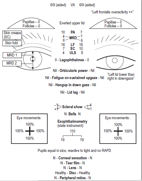 MRD gives more information than palpebral aperture (PA), the vertical distance between the upper and lower eyelid margins.
For example, a patient with unilateral ctropion and ptosis may have a similar PA to the normal side.
Levator function (LF) Typically 15–20 mm in the adult.
Block compensatory frontalis activity when measuring LF. Hold
a ruler vertically before the eye, between thumb and first finger. The second and third fingers rest firmly on the brow to overcome frontalis activity.
- Measure the maximal vertical excursion of the upper lid, from down gaze to up gaze.
- Note relative lid height in down-gaze. In congenital levator dystrophy the lid is higher than the fellow. In acquired ptosis (aponeurotic dehiscence) the lid is lower. Skin crease (SC) 7–8 mm in men, 9–10 mm in women.
- Marks the fulcrum of activity of the levator palpebrae muscle on the eyelid, and is formed where levator fibers attach to the skin.
- Lagophthalmos Residual inter-palpebral distance with gentle closure. Ask if there is nocturnal lagophthalmos.
- Scleral show (SS) Distance between the lid margin and the superior, and inferior, limbi, with each eye fixating the target in turn.
- Hang up in down gaze (failure of upper lid to descend normally). May occur with levator dystrophy, previous ptosis surgery, thyroid eye disease and orbital disease.
Additional observations
- Lid lag (phase lag on down gaze – a dynamic process).
- Cogan’s twitch (overshoot of upper lid on elevation from depression).
- Jaw movement (check for abnormal eyelid movement due to medial or lateral pterygoid synkinesis).
- Pupil reactions and size (check photopic and scotopic measurement if Horner’s syndrome is suspected).
- Saccades and ductions (may be affected with myopathy or aberrant third nerve regeneration).
- Bell’s phenomenon, corneal sensation (risk of corneal exposure following ptosis surgery if these are reduced).
- Take care to assess with each eye fixating in turn (e.g. apparent ptosis may be a pseudoptosis in the presence of hypotropia, or a double elevator palsy)
